Open 7 days
- Mon - Fri 8am-8pm
- Sat 8am-12pm
- Sun 8am-12pm
After hours veterinary care
After hours veterinary care
Mast cells are very nasty cancers in dogs seen frequently on the skin. They have many varying forms. We strongly advise you have all skin lumps checked that you find on your dog or cat. Often they aren’t anything to worry about but you must have them checked by a veterinarian.
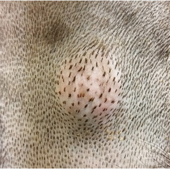
Mast Cell Tumours vary considerably in appearance however they are usually, round to oval, raised, pink to red, sometimes fleshy but sometimes merely a pink thickening of the skin. They sometimes don’t seem like much to worry about at all but they can be deadly.
Here is an example of a canine MCT
A good veterinarian will examine any unusual lesions for you and do a fine needle aspirate of the lump. The tumour cells can be seen in the cytology of the smear viewed under the microscope.
The mast cell cancer cells can also vary considerably depending on the grade of cancer. A feature of these cells is they contain lots of small granules of histamine. See the photo below. The large circle is the nucleus of the cell. The small purple dots are granules in the cytoplasm that contain histamine and also heparin.
Note the tiny purple granules in the cytoplasm of the cell. These are a feature of mast cell tumours.
If a diagnosis of canine MCT is made we will recommend staging the tumour with a view to wide margin surgical excision of the mass. We do surgery with curative intent in mind. The best chance of curing canine MCT is at the initial surgery with wide and deep surgical margins to make sure all of the tumour is removed with a healthy margin of normal tissue around it and below it.
Prior to aggressive curative intent surgery, it is important to stage the tumour. What does this mean exactly. Explained very simply, staging the tumour is an attempt to see whether it has spread already. Surgical removal is then important and the mass must be sent for histopathology for grading.
What is staging of canine mast cell tumours in the dog? Staging canine MCT in the dog is explained simply like this.
Once staging is done, we can determine whether surgery is going to be helpful. As mentioned previously, it is important to surgically remove these cancers with wide margins. The lesion is then sent for histopathology to confirm diagnosis and to grade the tumour. What is grading of canine mast cell tumours in the dog?
This is a simplified answer to help you understand what the different grades mean.
Explained in more scientific detail, a Grade 1 tumour shows well differentiated cells. They are contained and less likely to spread. Grade 2 MCT’s are less well differentiated and more likely to spread into adjacent cutaneous lymphatics and therefore find there way into other parts of the body. Grade 3 MCT’s are poorly differentiated or undifferentiated and are highly malignant, ie very likely to spread.
So if you see anything on your dog that looks like this (see below), you must go to the vet immediately and have it tested. Good outcomes can be achieved with diligent examination of your pet every month, accurate diagnosis, and good medical and surgical practices from your veterinarian. Canine MCT is a killer. You need to get any strange lumps tested by your vet.
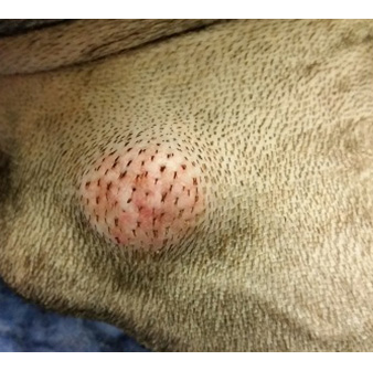
A feature of mast cell tumours is that they sometimes “degranulate” when the veterinarian does a fine needle biopsy. Remember that these tumours contain histamine and also heparin. Histamine can cause inflammation like redness of the tumour and adjacent skin and the heparin can cause a loss of coagulation causing bleeding.
This canine skin tumour was investigated using a FNA needle biopsy. Note the redness of the tumour and the area of haemorrhage that continued to bleed profusely for several minutes. This feature of canine MCT can create problems during surgical removal. Just occasionally we can run into problems with a lack of clotting and profuse haemorrhage following excision of the cancer. This is a recognised complication. It can sometimes be challenging to control bleeding after removal of canine MCT’s.
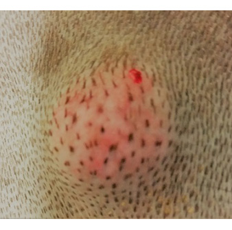
This is what this tumour looked like cytologically under the microscope.
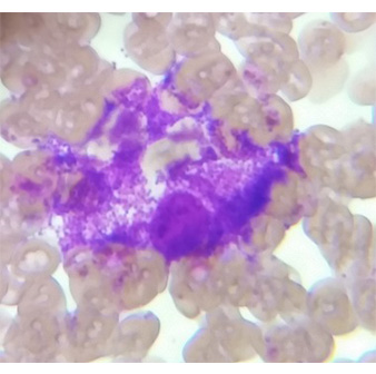
Note the cell membrane has ruptured and all the purple granules have escaped from the cells. This is called degranulation and causes redness, swelling and bleeding.
This is a crucial point. Owners often note that these tumours sometimes “swell up” but then go back down. The client becomes very worried when the tumour enlarged but then stops worrying when it goes back down so doesn’t go to the vet. If you notice a pink or fleshy lump on your dog that suddenly got bigger but then went back down, this is a feature of canine mast cell tumours in dogs. You must get it checked by your vet
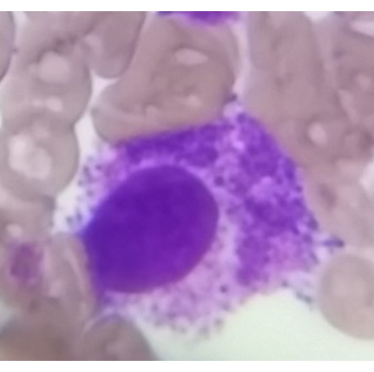
Here is another MCT. Note the large purple nucleus and purple granules.
What can I do if my dog has Grade 2 or Grade 3 canine mast cell tumours? Following surgical removal of the tumour chemotherapy is recommended.
What can I do if my dog has Grade 2 or Grade 3 canine mast cell tumours and I don’t want to do chemotherapy? You can palliate your dog with prednisolone. This may slow down the growth and spread of the cancer. It isn’t curative but may give you some extended time with your dog and improve the quality of life in the short term. For more information, visit Bunbury vets today!
Social distancing is easy for pets and people in our spacious waiting areas.


